Fig. 1.
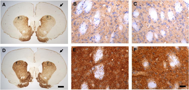
Representative tyrosine hydroxylase (TH) immunohistochemical staining of coronal sections from the striatum at 28 days after traumatic brain injury (TBI; D, E & F) revealed a general increase in TH immunoreactivity (TH-IR) with a dense plexus of immunoreactive fibers and punctate vesicles compared to the sham controls (A, B & C). The increase of TH-IR is bilateral but more evident in the contralateral striatum (B & E) compared to the ipsilateral striatum (C & F). A (sham) and D (TBI) are the lower magnified microscopic view (1 × objective lens) of TH immunostaining. B & C (sham) and E & F (TBI) are the sister sections of A and D in the high magnified microscopic view (40 × DIC objective lens) of TH immunostaining counterstained with hematoxylin. B & C and E & F are the close-up views of the areas indicated in panels A and D. Arrow Point: ipsilateral side (in A & D). Bars: A = D = 1,000 μm (1 mm); B = C = E = F = 25 μm.

