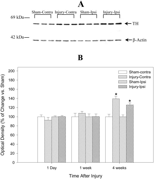
Fig. 3.
Summarizes the semiquantitative measurements of tyrosine hydroxylase (TH) protein in rat striatum after traumatic brain injury (TBI) or sham operation. The lower Panel (B) is the histograms of Western blot data from the ipsilateral (Ipsi) and the contralateral (Contra) striatum homogenates at 1 day, 7 days, and 28 days after TBI or sham operation and demonstrates that TH protein is increased bilaterally but more evident in the contralateral side at 28 days after TBI (N = 6, * = p < 0.05). The upper panel (A) is the representative gel bands (N = 3) at 28 days after TBI or sham operation. There is no significant statistical difference in TH optical densities with or without normalizing to β-actin. The number on left of immunoblot indicates the position of the molecular weight marker (A).

