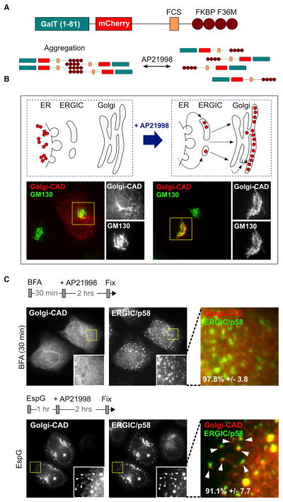
Figure 1. EspG Does Not Interfere with Protein Export from the ER.
(A) Schematic representation of the Golgi-CAD construct design. Aggregation of Golgi-CAD is mediated by FKBP F36M domains and can be reversed by addition of AP21998 molecule.
(B) Development and confirmation of the inducible ER-to-Golgi trafficking assay to study protein transport through the early secretory pathway. The trans-Golgi-associated marker (Golgi-CAD) is trapped in the ER upon transfection (left), but is trafficked to the Golgi after the addition of the small molecule AP21998 (right).
(C) Fluorescent micrographs show the final localization of Golgi-CAD 2 hr after addition of AP21998. Golgi-CAD escapes the ER but is arrested near p58-positive clusters (arrowheads) in cells microinjected with EspG (asterisks). No ER export occurs in cells treated with BFA.
See also Figure S1.

