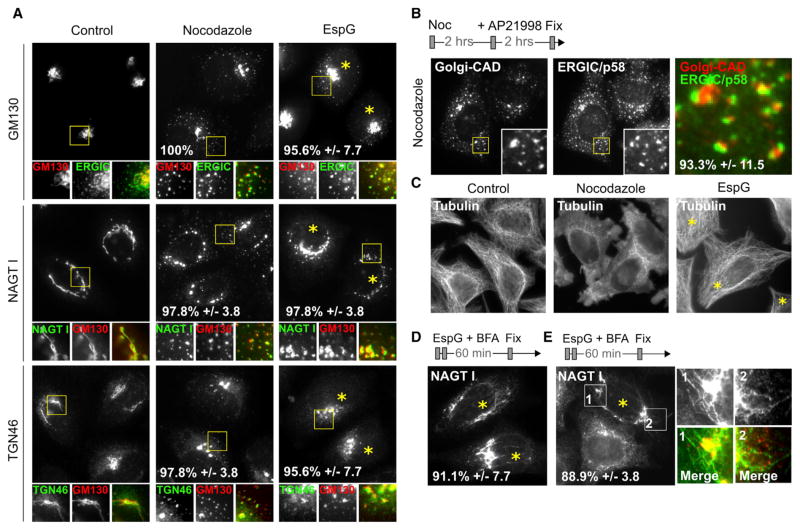
Figure 2. EspG Functionally Mimics Disruption of Microtubule-Dependent Processes.
(A) Fluorescent micrographs show Golgi morphology relative to positioning of cis- (GM130), medial- (NAGT I), and trans-Golgi (TGN46) markers in cells treated with nocodazole or microinjected with EspG.
(B) Nocodazole prevents Golgi-CAD transport at p58-positive clusters during the inducible trafficking assay in a manner analogous to that observed for EspG.
(C) EspG shows no apparent defect in microtubule network, despite the similar presentation of Golgi phenotypes. Microinjected cells are marked with an asterisk.
(D) BFA-induced Golgi tubules persist in EspG-injected cells and fail to fuse with ER, suggesting inhibition of membrane transport along microtubules. Microinjected cells are marked with an asterisk.
(E) Persistent BFA-induced Golgi tubules (green) emerge from and connect cis-Golgi (GM130, red) in cells transfected with EspG (asterisks).
See also Figure S3.

