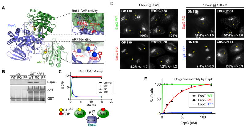
Figure 3. Separating the Roles of ARF1 and Rab1 in the EspG Mechanism.
(A) Identification of predicted ARF1- and Rab1-specific functional mutants based on structural data of EspG bound to ARF and Rab GTPases (3PCR, 4FMC, and 4FME). Mutations are shown in red, and blue spheres denote water molecules.
(B) Pull-down experiments testing the ability of EspG mutants to bind ARF1.
(C) Rab1-GAP assay testing the GAP activity of EspG constructs.
(D) Golgi disassembly by EspG deficient for either ARF1 binding or Rab1-GAP activity as determined by Golgi ribbon fragmentation and enlarged p58 clusters. Representative micrographs are shown for the lowest and highest concentrations tested. Microinjected cells are marked with an asterisk.
(E) Quantification of disrupted Golgi and p58 clusters phenotype. Needle concentrations are shown.
See also Figures S2 and S3.

