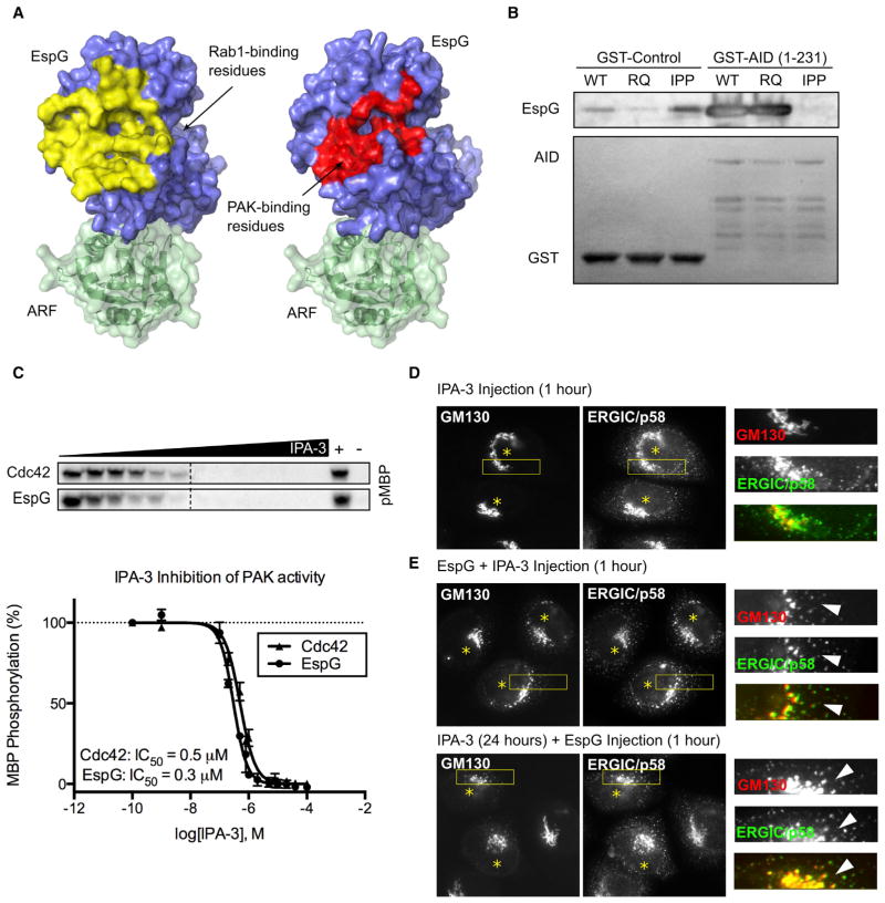
Figure 4. Fragmentation of the Golgi by EspG Is PAK Independent.
(A) PAK and Rab1 share the binding surface on EspG. Protein Data Bank structures 3PCS and 4FME were used for alignment.
(B) Binding of EspG constructs to PAK AID. The ARF1-binding deficient mutant (IPP) does not interact with PAK.
(C) Kinase assay showing the sensitivity of PAK activation to IPA-3. Radiography data from the assays were quantified and plotted to determine IC50 values.
(D and E) Golgi fragmentation is induced by EspG in cells treated with IPA-3 to inhibit PAK. Microinjected cells are marked with an asterisk.

