
Figure 3.
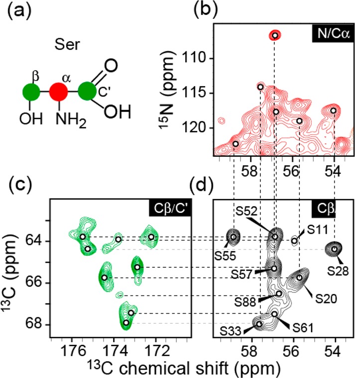
Identification of serine residues of ΔN6 fibrils using 2D MAS NMR and variously labeled samples. (a) 13C-labeling scheme of serine using [2-13C]-glycerol (red) or [1,3-13C]-glycerol (green) as the carbon source.74,76 (b) One-bond ZF-TEDOR of [2-13C-glycerol]-ΔN6. (c) Multibond RFDR of [1,3-13C-glycerol]-ΔN6 using an 11 ms mixing period. (d) One-bond RFDR of U–13C, 15N-labeled ΔN6 recorded using a 1.6 ms mixing period. Spin systems of all nine serine residues were identified by their characteristic downfield Cα and Cβ chemical shifts. The assignments were from the following 2D and 3D spectra. Dashed lines guide the assignment of each residue.