
Figure 6.
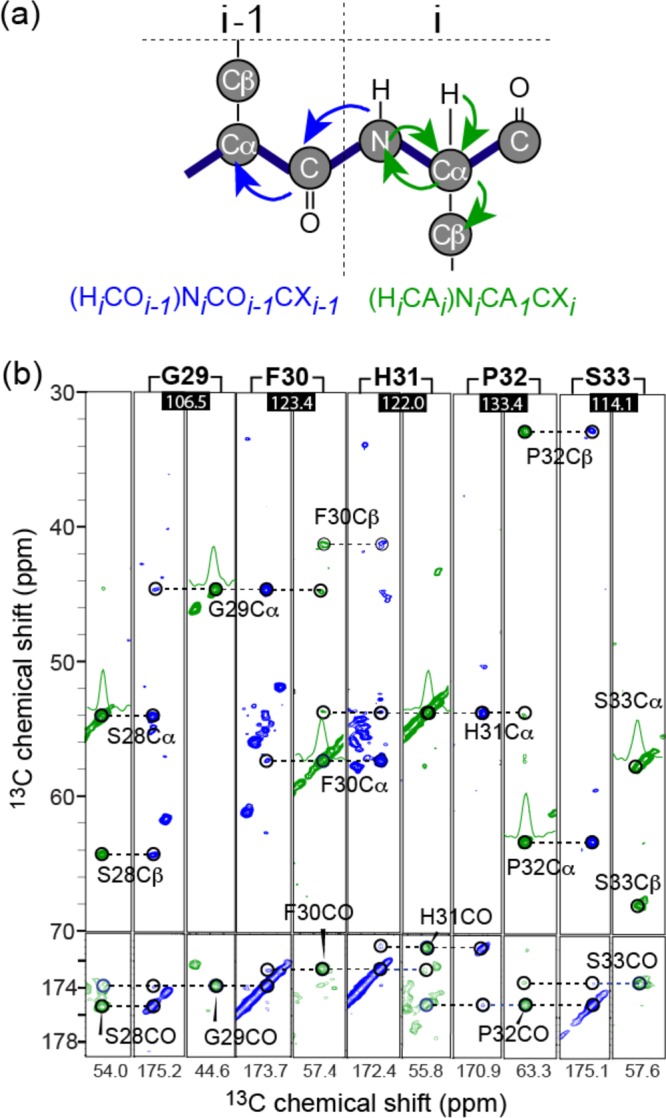
Representative sequential backbone walks from S28 to S33 in 3D TEDOR-CC spectra of ΔN6 fibrils. (a) Simultaneous transfers of N(i)–C′(i – 1) and N(i)–Cα(i). The initial magnetization in TEDOR-CC is from 13C–1H CP, in contrast to the 15N–1H CP in conventional 3D 15N–13C–13C experiments, providing the optimal enhancement of proline intensity. (b) 2D 13C–13C (F1–F3) planes of the 3D TEDOR-CC spectrum of ΔN6 fibrils. 15N chemical shift (F2) for each 2D plane is indicated in black squares. CO and Cα chemical shifts are shown on the x-axis. One-dimensional cross sections are shown for Cα peaks in NCACX spectra in green. Homonuclear 13C–13C mixing was accomplished using 4.8 ms RFDR. The spectra were acquired using the U-ΔN6 fibril on a 900 MHz spectrometer (1H frequency).