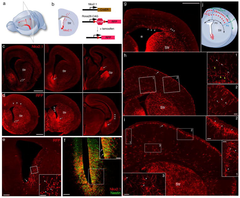
Fig. 1.
Nkx2.1+ progenitors in the embryonic VGZ are a novel source of cortical interneurons. (A) NKX2.1 expression (red) in middle-caudal regions of the late embryonic VGZ. Arrows indicate the migration of VGZ-derived cells toward the dorsal and rostral cortex. (B) A coronal section from a region indicated in (A). Red dots represent VGZ-derived cells migrating toward the cortex. Genetic fate mapping strategy is depicted to the right. In an Nkx2.1CreER;Ai9 mouse, Cre-mediated recombination is induced by tamoxifen and activates RFP expression. Str, striatum. (C) Immunofluorescence showing NKX2.1 protein (arrows) in the middle-to-caudal region (middle and right panels) but not rostral region (open arrow in left panel) of E17 VGZ. (D) Nkx2.1CreER;Ai9 mice induced at E17 and examined at E18. RFP-labeled progenitors were abundant in the middle-caudal regions (solid arrows) but not rostral region (open arrow) of VGZ. RFP+ migrating cells (arrowheads) were found not only in midcaudal sections but also in the rostral section. (E) Coronal section of an E18 brain induced by low-dose tamoxifen. More sparse VGZ progenitors (arrow) and postmitotic cells (arrowheads in inset) are labeled, giving a clearer view of the migratory stream along the lateral wall of the ventricle. (F) Colocalization (arrow) of NKX2.1 and Nestin in VGZ progenitors shown by double immunostaining. (G) Coronal section of a P0 brain induced at E17. Migrating cells emerge from the dorso-lateral wall of the ventricle (star) and split into lateral and medial stream (arrowheads) within the cortical subventricular zone. (H) Coronal section of a P2 brain induced at E17. Migrating cells, each with a characteristic leading process (arrowheads in insets) pass through cortical plate into L1 (arrow). (Insets) Tangentially migrating cells in the subventricular and intermediate zone (yellow arrowheads, H1) and radially migrating cells in the cortical plate (light gray arrowheads, H2). (I) Coronal section of a P7 brain induced at E17. RFP+ cells descend from L1 into the cortex and settle at the L1/L2 border (arrow), forming dense clusters, and start to differentiate (arrowheads in I1 and I2). Deep-layer RFP+ cells (arrowheads in I3) also settle and show sign of differentiation by this stage but their migration route is unclear. (J) Diagram depicting the migration route and schedule of VGZ-derived cortical cells. Scale bars: 500 μm in (C), (D), and (G to I); 200 μm in (E); 50 μm in (F) and insets of (E), (H), and (I); and 25 μm in inset of (F).

