
Fig. 2.
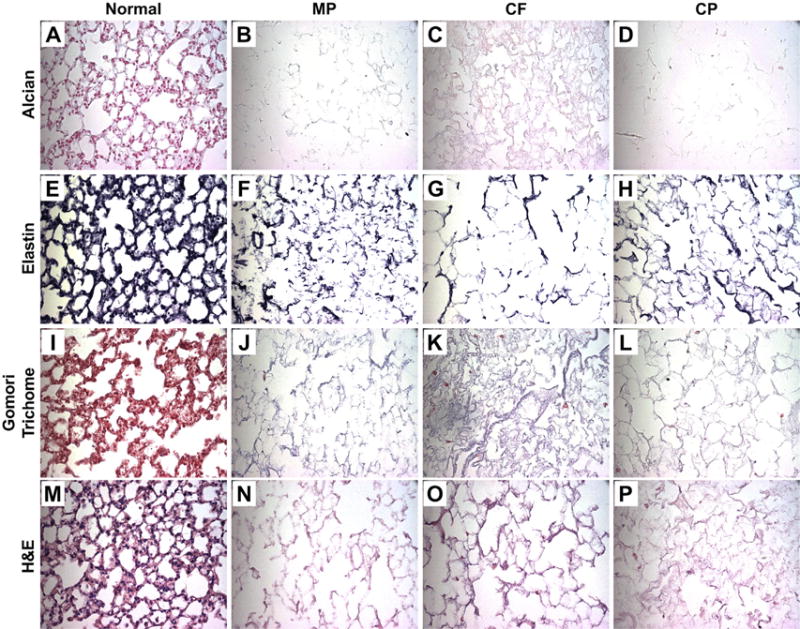
Histologic Evaluation of De-cellularized Scaffolds Representative images for Alcian Blue, a glycosaminoglycan's stain, demonstrate a loss across all three de-cellularization methods (blue) (A–D). Verhoeff's elastin stain, which outlines elastin filaments (black) (E–H), also demonstrates a loss across all three methods. Gomori Trichrome, a collagen stain (blue) (I–L) also indicates a loss across all three methods. Lastly, H&E staining (M–P) confirms loss of nuclei, while maintain structural integrity in all three methods. Representative images are shown from triplicate lungs de-cellularized using each method (All Images 40× Magnification). (For interpretation of the references to colour in this figure legend, the reader is referred to the web version of this article.)