
Fig. 4.
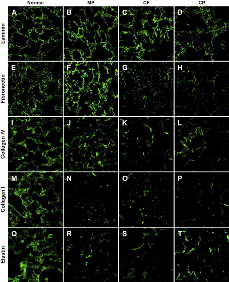
Qualitative Analysis of ECM Proteins by Immunofluorescence Representative images from triplicate lungs de-cellularized using each method and immunostained for laminin (A–D), fibronectin (E–H), collagen IV (I–L), collagen I (M–P), elastin (Q–T) qualitatively demonstrate loss of ECM proteins from the de-cellularization process. The MP protocol appears to retain the most laminin, fibronectin and Collagen IV compared to the CF and CP protocols. The CP method appears to retain the most elastin. (All Images 63× Oil Magnification).