
Fig. 6.
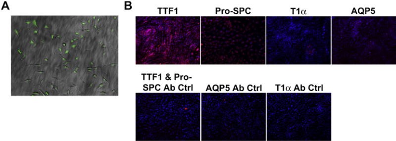
Characterization of C10 Cells After GFP Transfection (A) C10 alveolar epithelial cells express GFP following viral transduction and FACS sorting. C10 cells following flow-based sorting were over 95% GFP positive (10× Magnification). (B) C10-GFP cells cultured on untreated plastic express type II alveolar epithelial markers TTF-1 and Pro-SPC, but do not express type I alveolar epithelial markers such as AQP5 or T1α (20× Magnification).