
Fig. 9.
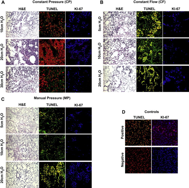
Histology, Apoptosis and Cell Proliferation of Re-cellularized Scaffolds After 3 Days In Culture Delivery pressure of maintenance medium was examined in range of 5–30 cm H2O (A) CP Seeded lungs appear to repopulate the scaffolds by H&E, but only yield proliferative cells when maintenance medium is delivered at a pressure of 20 cm H2O. (B) CF seeded lungs also appear to repopulate the scaffolds by H&E; however, all cells display an apoptotic, non-proliferative phenotype under all perfusion pressure conditions assessed. (C) MP seeded lungs were also apoptotic and non-proliferative under all perfusion pressure conditions assessed. (D) TUNEL negative control was normal lung and TUNEL Positive control was normal lung treated with DNAse (20× magnification). Ki67 negative control was no primary; positive control was C10-GFP cells dividing in culture (20× magnification). Ki67 staining (TOPRO3 = BLUE, Ki67 = RED), TUNEL Staining (PI (Nuclear Counterstain) = RED, TUNEL = Green, CELL DEATH = Yellow). (For interpretation of the references to colour in this figure legend, the reader is referred to the web version of this article.)