Fig. 2.
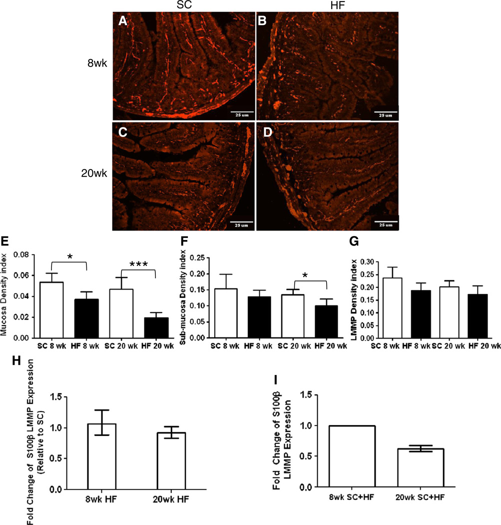
Effect of diet on S100β expression in mouse duodenum. 10 μmduodenum cryostat sections of SC (A, C) and HF (B, D) mice were stained with antibodies to S100β protein after 8 (A–B) and 20 weeks (C–D). Analysis of density indexes (stained area/total tissue area) in the mucosa showed a significant decline in HF diet mice after 8 and 20 weeks (E; 8 weeks, n = 5, *P < 0.05; 20 weeks, n = 6–7, ***P < 0.001; unpaired students t-test). EGC density index of sub-mucosal plexus showed a decline due to high-fat diet only after 20 weeks (F; 8 weeks, n = 5; 20 weeks, n = 6–7, *P < 0.05; unpaired students t-test) while density in LMMP remained unchanged (G; SC n = 5; HF n = 6–7). RT-PCR expression levels of S100β in LMMP of HF diet fed mice was not significantly different than SC (H; 8 weeks n = 4; 20 weeks, n = 6), and was also not significantly different based on mouse age (I; n = 4).

