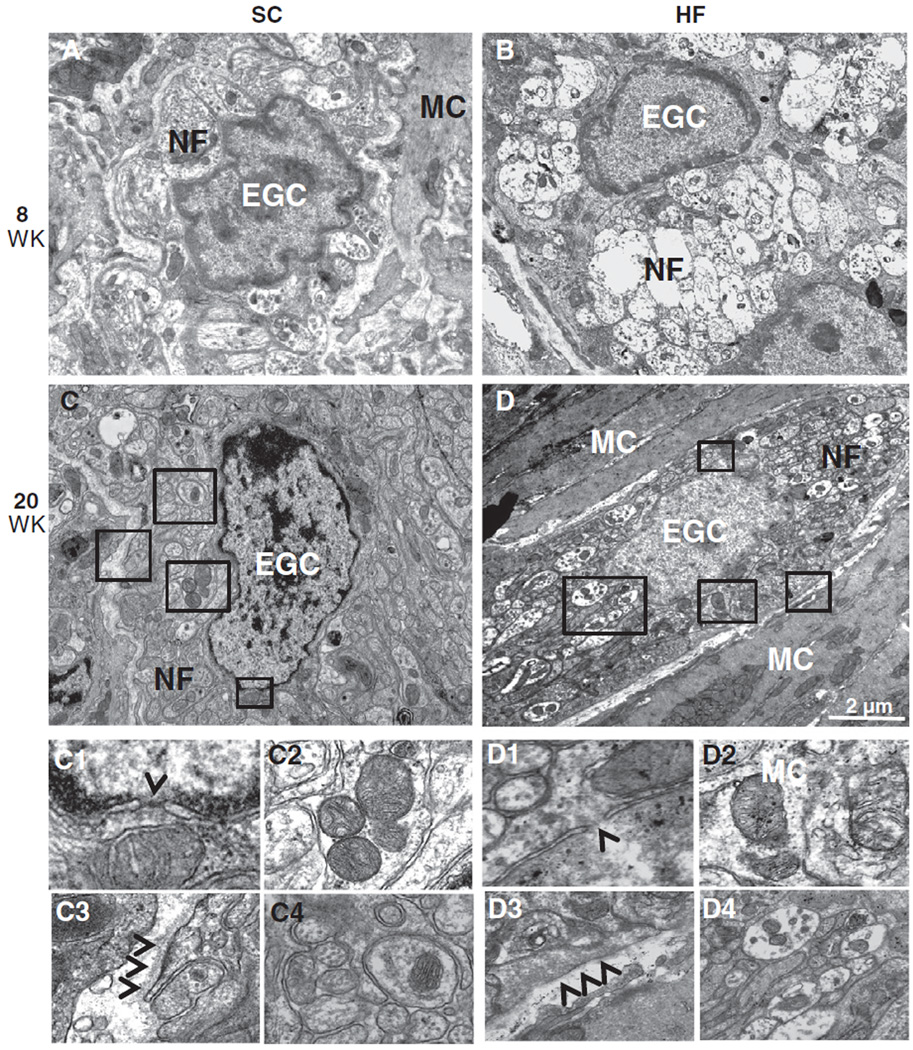
Fig. 5.
TEM analysis of EGCs in the myenteric plexus did not show differences between SC and HF mice. Ultrastructural analysis of EGCs revealed normal morphology in SC (A, C) and HF (B, D) mice at 8 and 20 weeks. Inserts show karyolemm and nuclear pores with no signs of rupture or apoptosis (arrowhead: C1 and D1), healthy mitochondria (C2 and D2) and no signs of basement membrane thickening (arrowheads: C3 and D3) around EGCs of each mouse group. This is regardless of the early signs of neuropathy (axonal edema and loss of neurofilaments) seen in unmyelinated nerve fibers (NF) of HF (D4) but not SC (C4) mice. Muscle cells (MC) are also seen and exhibit normal morphology.

