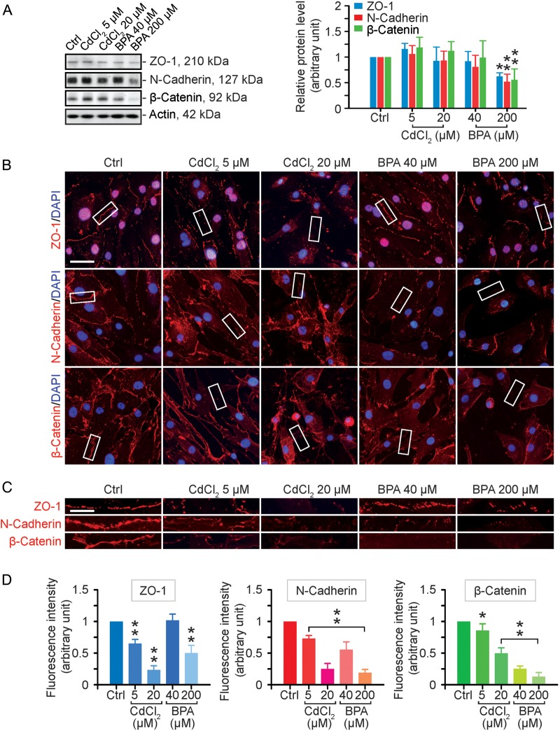
Figure 1.
A study to assess the effects of cadmium chloride (CdCl2) and bisphenol A (BPA) on the expression and localization of cell adhesion proteins in human Sertoli cells. (A) Human Sertoli cells were cultured on fibronectin-coated dishes and exposed to CdCl2 (5 and 20 µM) or BPA (40 and 200 µM) for 2 days. Thereafter, cells were harvested for lysate preparation and for immunoblotting (left panel). The histogram in the right panel summarizes the immunoblotting data. Protein bands were densitometrically scanned, and normalized against β-actin; each bar is a mean ± SD of n = 3 independent experiments. Controls were normalized to 1.0. *P < 0.05; **P < 0.01 with treatment versus corresponding control (Ctrl) group. (B) Immunofluorescence analysis of some blood–testis barrier (BTB)-associated proteins (all in red fluorescence): zonula occludens-1 (ZO-1, a tight junction adaptor), N-cadherin (a basal ectoplasmic specialization (ES) integral membrane protein) and β-catenin (a basal ES adaptor). Cell nuclei were visualized by DAPI (4′,6-diamidino-2-phenylindole) staining. Scale bar = 40 µm. (C) Enlarged images corresponding to the white boxes in (B) of ZO-1, N-cadherin and β-catenin at the human Sertoli cell–cell interface, illustrating changes in their localization following exposure to toxicants. Scale bar = 15 µm. (D) Image analysis of the changes in the fluorescence intensity of cell adhesion proteins at the human Sertoli cell–cell interface. Each bar is a mean ± SD of three experiments including ∼100 cells per experiment. Controls were normalized to 1.0 in each experiment. *P < 0.05; **P < 0.01 with treatment versus corresponding control (Ctrl) group.

