
Figure 7.
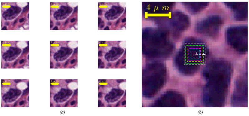
(a) Images of a CB cell created based on different points inside its body. The middle image is the image created based on the pathologist’s marking. (b) Image of a cell used on the examination of COB and CLEM’s consistency. New images of the cell were created based on pixels that lie on the colorful rectangulars shown in the figure (created based on a distance d from the cell’s center, shown with a white arrow).