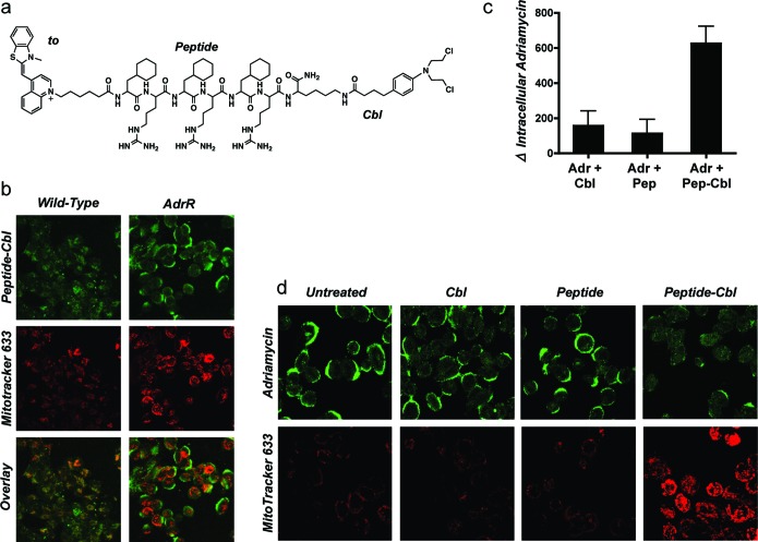
Figure 1.
Peptide-Cbl conjugate modulates Pgp activity. (a) Structure of fluorescently labeled peptide-Cbl conjugate used in these studies. Thiazole orange (to) was used for fluorescent labeling of the peptide-Cbl conjugates. The peptide was an alternating array of d-arginine (r) and cyclohexylalanine (Fx). (b) Intracellular localization of 5 μM peptide-Cbl (green), MitoTracker 633 (red), and signal overlay in live wild-type and adriamycin-resistant A2780 cells. (c) Changes in intracellular levels of adriamycin assessed by flow cytometry in cells overexpressing the Pgp efflux pump. All inhibitors were tested at 5 μM. Mean values are plotted, n = 3; error bars are SEM. See Figure S1 in the Supporting Information for basal levels of uptake. (d) Intracellular localization and uptake of adriamycin and MitoTracker 633 as observed by confocal microscopy. All inhibitors were tested at 5 μM. Detector voltages were kept constant for each panel of images to allow for a comparison of signal intensities. Results show that neither peptide nor Cbl alone is sufficient to inhibit Pgp activity, and both components of the peptide-Cbl conjugate are required.

