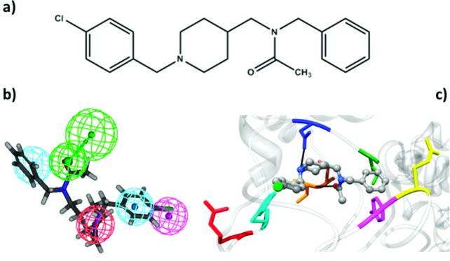
Figure 3.

(a) Structure of compound EL-1 designed exploiting the 2D/3D interaction model and the 3D homology model of the σ1 receptor developed in this work. (b) Mapping of EL-1 onto our 3D pharmacophore model developed for σ1 ligands.14−14d (c) Modeled complex of the σ1 receptor with EL-1 showing the key interactions proposed in the topographical interaction model depicted in Figures 1b and 3b. The main protein residues involved in these interactions are Arg119 (red), Trp121 (cyan), Asp126 (blue), Ile128 (forest green), Thr151 (sienna), Val152 (orange), Glu172 (yellow), and Tyr173 (magenta).
