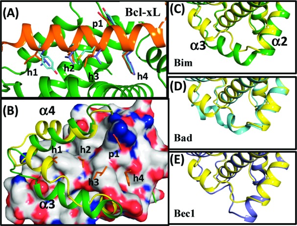
Figure 1.

(A) Superimposition of Bcl-xL with the Bim (orange), Bad(cyan), and Bec1 (blue) BH3 peptides. (B) Structural alignment between the ligand-free Bcl-xL(yellow) and Bcl-xL with the Bim peptide(green). H1-4 and p1 residues of the Bim peptide are shown in orange. Conformational changes of the α3 helix in Bcl-xL when binding to (C) Bim (green), (D) Bad (cyan), and (E) Bec1 (blue) peptide. The reference structure of ligand-free Bcl-xL is shown in yellow. The PyMOL program (www.pymol.org) was used to view the protein models and prepare the graphics.
