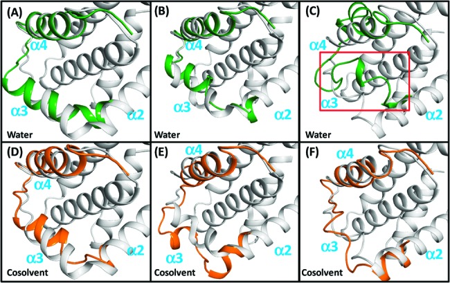
Figure 2.

Comparison of the crystal structures of the Bim-bound (A, D), the Bad-bound (B, E), and the Bec1-bound (C, F) Bcl-xL with snapshots of conformations after 32 ns simulations. Conformations of crystal structures; snapshots in water and in cosolvent are colored in gray, green, and orange. Only residues F97−N136 in the snapshots are shown for clarity.
