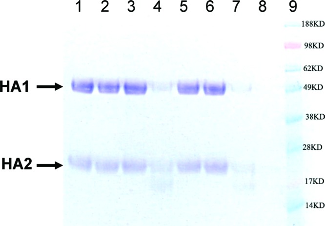
Figure 2.

SDS-PAGE analysis of trypsin sensitivity assay showing 1 and 40 protected purified HA from trypsin digestion. Lane 1, purified HA; lane 2, HA treated with trypsin without a prior acidification step; lane 3, acidified HA without trypin treatment; lane 4, DMSO control; lane 5, compound 1 (10 μM); lane 6, compound 40 (10 μM); lane 7, ribavirin (10 μM); lane 8, trypsin only; and lane 9, molecular markers. Lanes 4–7 included all steps and components in a typical trypsin sensitivity assay. Positions of HA1 and HA2 are marked.
