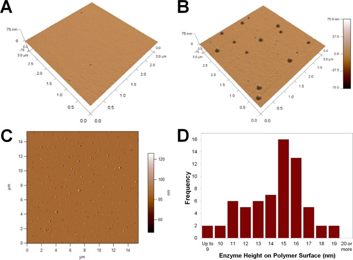
Figure 1.
Tilted view of a 3 μm × 3 μm AFM scan of a PMMA surface following UV activation and incubation with 7 μg/mL λ-Exo enzyme without (A) and with (B) EDC/NHS coupling reagents. (C) A 15 × 15 μm phase image of a PMMA surface incubated with 7 μg/mL λ-Exo enzyme with EDC/NHS coupling. Surface AFM analysis revealed an RMS roughness of 1.58 ± 0.18 nm. (D) Histogram of the height of features on the activated PMMA surface and subsequently functionalized with λ-Exo determined by taking an AFM line scan across each immobilized enzyme and measuring the maximum height of the feature.

