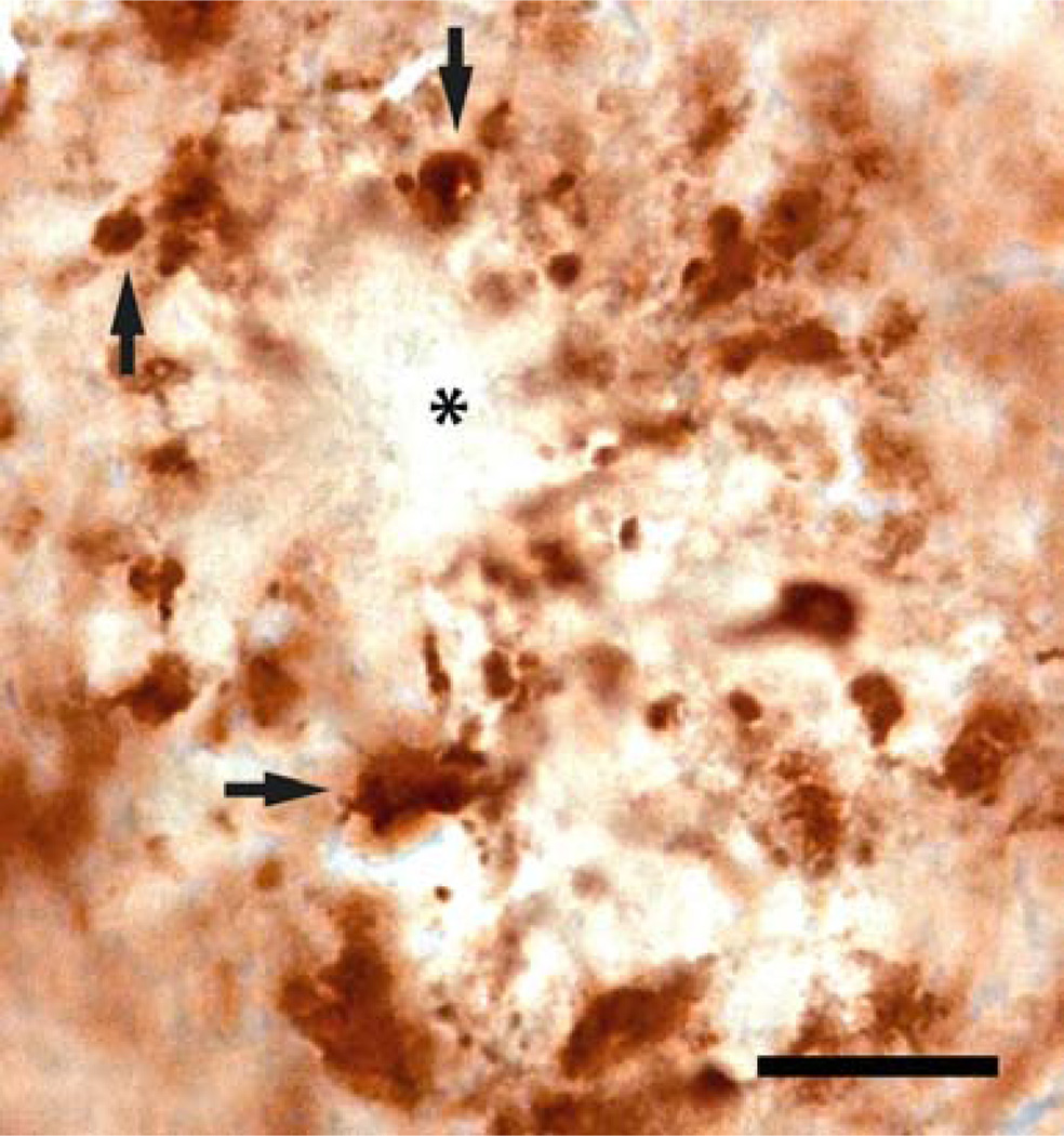
Figure 8.
High magnification of synaptophysin immunoreactivity in the hippocampus of a 10-month-old APPSL/PS1ho KI mouse, showing the diminished synaptophysin signal from the core of an amyloid deposit (asterisk). Note the enlarged SIPBs (synaptophysin-immunoreactive presynaptic boutons) surrounding the plaque (black arrows). The scale bar represents 300 µm.

