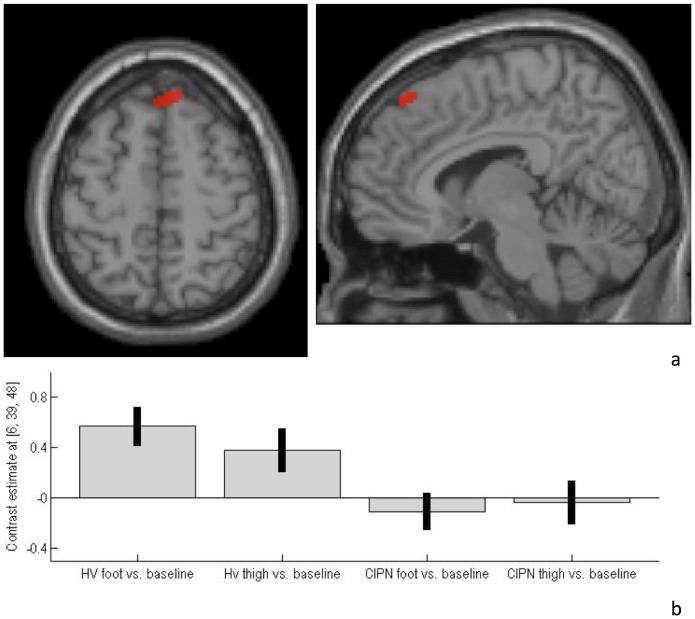
Figure 1. CIPN-myeloma patients demonstrated a) hypo-activation of superior frontal gyrus during heat-pain stimulation, compared with healthy volunteers.
Functional imaging data are shown overlaid on both axial (z = 48 mm) and sagittal (x = 6 mm) slices through a canonical single-subject T1-weighted image. For display purposes, the statistical threshold is p<0.001, uncorrected, at the voxel-level. b) Contrast estimates and 90% CI at co-ordinate 6, 39, 48 for both healthy volunteer and CIPN-myeloma patient groups.

