
Figure 2.
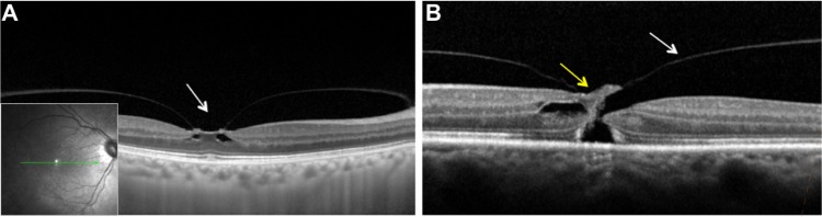
Images of macular region on spectral domain optical coherence tomography (SD-OCT; Spectralis OCT, Heidelberg Engeneering, Heidelberg, Germany).
Notes: (A) Vitreomacular traction with focal traction at foveal (arrow) region and intrafoveal pseudocysts. (B) Full-thickness macular hole with vitreomacular traction (yellow and white arrows, respectively).