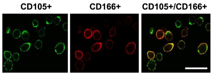
Figure 1. Immunocytochemical analyses of cells isolated from articular cartilage of OA subjects stained with CD105-PE and CD166-FITC.

Samples were collected from 5 normal subjects and 10 OA patients. Data from 3 independent experiments were combined. Fluorescence was examined using a confocal laser microscope. Scale bar represents 50 µm.
