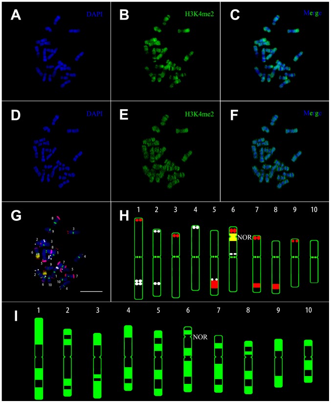
Figure 1. Karyotyping of immunolabelled metaphase chromosomes.
(A-C) Original image of chromosomal distribution of H3K4me2 from a maize inbred line (chang7-2) (2D). (D-F) Enhancement of the contrast of the original image by three-dimensional reconstruction after deconvolution (3D). (A, D) 4, 6-diamidino-2-phenylindole (DAPI) staining signals in blue, (B, E) immunostaining signals in green and (C, F) merge of both. (G, H) FISH karyogram constructed from the same spread. Assignments of pseudo-colors to each probe: TAG as white, CentC as green, 45s rDNA as yellow and knob 180-bp as red. (G) Metaphase chromosome identification by combining the four probes and DAPI staining. (H) Ideogram of FISH karyotype indicating the position of the four probes. (I) Karyogram of the H3K4me2 profile constructed from the immunolabelled chromosomes by three-dimensional reconstruction after deconvolution. NOR: nucleolus organizing regions. Scale bar = 10 µm.

