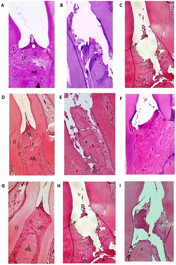
Figure 3. Microscopic analyses.
A, D and G: Normal periodontium. B, E, H: Periodontium from a rat with periodontitis (treated with saline) showing alveolar bone and cementum resorption (discontinuous cementum) and inflammatory cell infiltration. C: Treatment with AZT (1 mg/kg) and I: Treatment with AZT (10 mg/kg) showing no reduced inflammation and increased alveolar bone loss. F: Periodontium from a rat with periodontitis (treated with AZT, 5 mg/kg) showing reduced inflammation and decreased alveolar bone loss. Sections were stained with H&E. Original magnification 40×. Scale bars = 100 µm. G, gingiva; PL, periodontal ligament; D, dentin; AB, alveolar bone; C, cementum; a, bone loss; b, resorption of cementum; c, inflammatory process; e, f, decreased inflammation process and bone loss.

