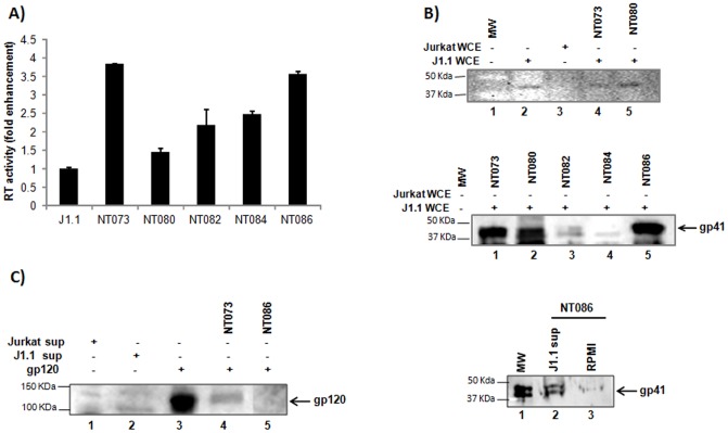
Figure 3. Capture of HIV-1 virions by nanotrap particles.
(A) A 30% slurry of each nanotrap particle in unsupplemented RPMI (30 µl) was incubated with J1.1 cell supernatants (200 µl) for 30 min at room temperature. Virus-bound nanoparticles were washed to remove unbound virus, and the nanoparticle pellets were suspended in 50 µl of Tris (250 nM) + 1% Triton X-100, and assayed for RT activity without elution. Fold enrichment of virus binding to nanotrap particles was analyzed over J1.1 supernatant alone without prior enrichment. (B) HIV-1 gp41 was detected in J1.1 WCE by WB without nanoparticles pretreatment (upper panel, lane 2). The uninfected Jurkat WCE was used as negative control (lane 3). Binding of HIV-1 by nanotrap particles through interaction with gp41 was detected in J1.1 WCE following treatments with nanoparticles NT073 and NT080 (lanes 4 and 5, respectively). The captures of gp41 from J1.1 WCE were also shown by nanoparticles NT073, NT080 and NT086 but not by NT084 (middle panel). Fresh antibody was used for this panel. (C) Interaction of nanotrap particles with HIV-1 envelope gp120 was detected by western blot using anti-HIV human serum. Briefly, purified gp120 glycoprotein (1 µg) or J1.1 culture supernatant was spiked into water and incubated with or without nanotrap particles slurry for 30 min at room temperature, washed and suspended in Laemmli buffer before resolved on SDS-PAGE gel and analyzed by western blot.

