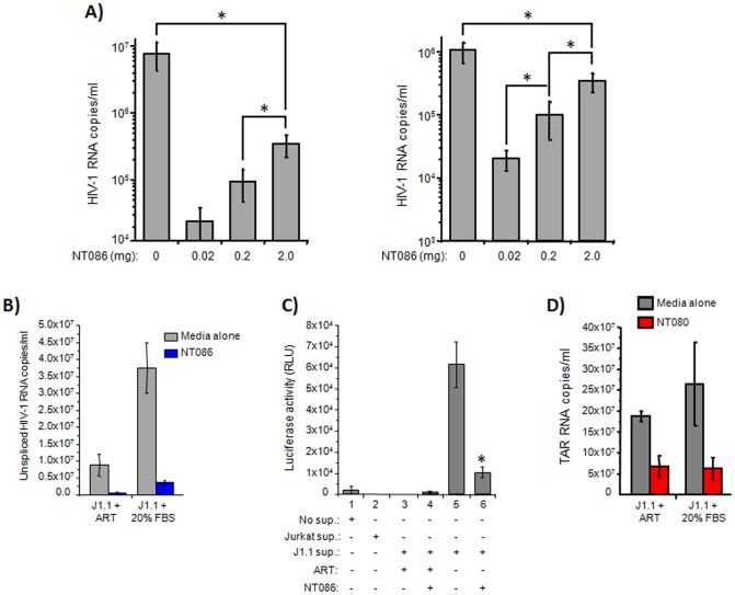
Figure 6. HIV-1 and exosomes capturing capacity of nanoparticles.
(A) 100 µl aliquot of the dual-tropic HIV-1 89.6 containing 10 or 1 ng p24/ml was incubated with a pellet of 0.02, 0.2 or 2.0 mg of NT086 for 30 min at room temperature. The virus bound-nanoparticles were washed and RNA isolated. The RNA was converted to cDNA and PCR reaction mixtures were prepared using high-low concentrations of cDNA, Gag primers and the iTaq Universal SYBR Green Supermix. Serial dilutions of DNA from 8E5 cells were used as the standards. Quantitative real-time PCR reactions were carried out in triplicate. The asterix represents the statistical significance at the level of p<0.05. (B) The infected J1.1 cells were treated with or without anti-retrovirals (ART), and then sups were incubated with NT086 to capture virions. After washing, the total RNA was extracted from the nanoparticle captured materials and then subjected to qRT-PCR using specific primers to unspliced HIV-1 RNA. (C) The infectivity of the nanoparticles captured virions were also analyzed by incubating them with TZM-bl cells and measuring luciferase activity. The asterix represents the statistical significance at the level of p<0.05. (D) The NT080 nanoparticles capture of exosomal RNA from J1.1 supernatants with or without pretreatment of ART were similarly measured by qRT-PCR using specific primers to TAR RNA.

