
Figure 4. 5-Methylcytidine (5-mC) immunostaining of placental samples.
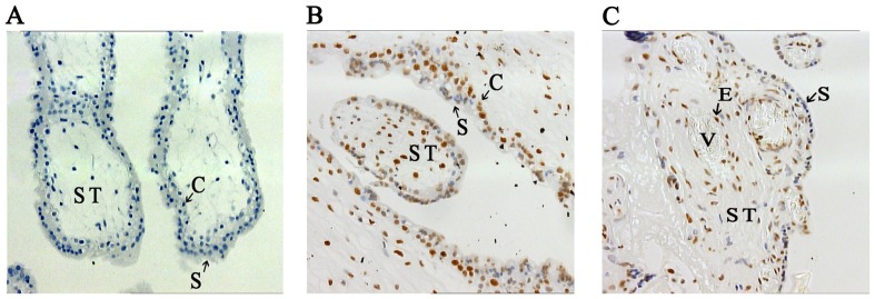
Tissue sections from paraffin-embedded placental tissues were treated with primary (Mouse anti 5-mC, concentration: 1/800), secondary (horse anti-mouse, concentration: 1/400) antibodies. Color development was performed with immunoperoxidase system. The 5-Methylcytidin-positive cells were stained brown, while negative cells displayed blue color of hematoxylin counterstaining. A. The negative control (1N) (20×10 magnitude) without primary antibody. B. 5-mC immunostaining of 1N placenta (20×10 magnitude). C. 5-mC immunostaining of 3N placenta (20×10 magnitude). Compared to 1N placenta, thinner lay of syncytium was observed in 3N placenta. Syncytiotrophoblasts (S) were stained relatively lighter than cytotrophoblast (C), stromal cells (ST), and epithelial (E) cells. V, blood vessels. Overall, similar intensity of 5-mC staining was observed 1N and 3N placentas.