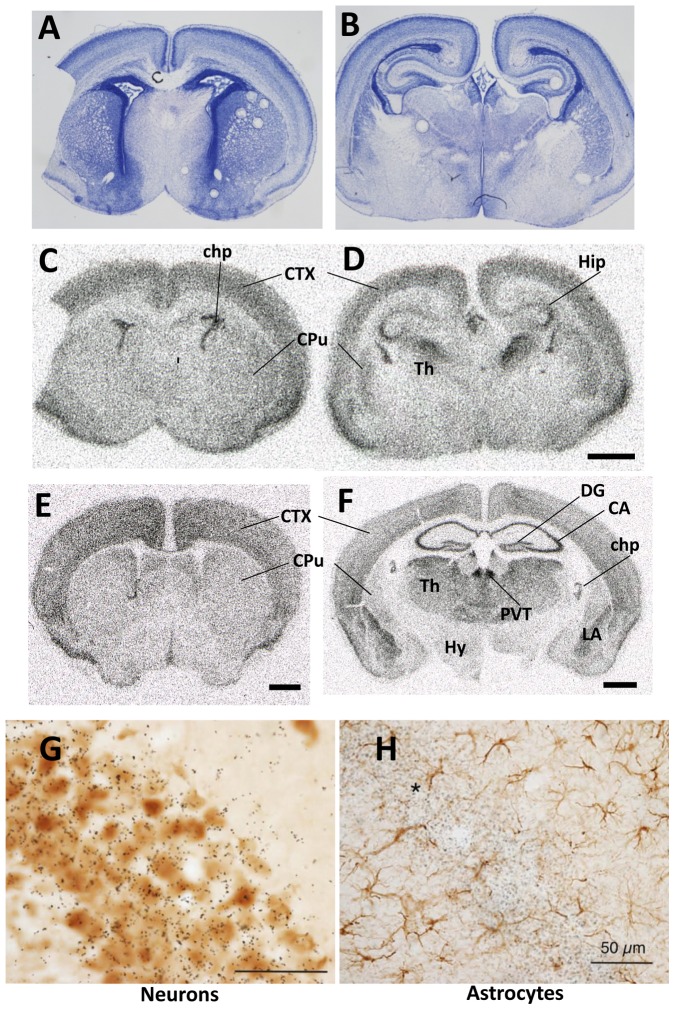
Figure 1. Lat2 expression.
A–D: P0: A and B are Nissl staining and C and D in situ hybridization radioautographs. E and F: P21 in situ hybridization radioautographs. Abbreviations: chp, choroid plexus; CTX, cerebral neocortex; CA, cornus Ammonis; CPu, caudate-putamen; DG, dentate gyrus; Hy, hypothalamus, Hip, hippocampus; Th, thalamus; PVT, thalamic paraventricular nucleus; LA, lateral amygdala. G: in situ hybridization (P21) with 35S-Lat2 probe combined with immunohistochemistry for NeuN to reveal neurons. Hippocampal CA1 field. H: Similar field as G, but at lower magnification, with cells stained for glial fibrillary acidic protein (GFAP). The majority of the silver grains can be seen on the neuronal pyramidal layer (asterisk) with background signal on the astrocytes. Scale bars were 1 mm in C–D, E and F, and 50 µm in G and H.

