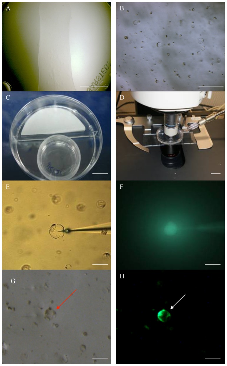
Figure 5. Microinjection of DNA into oil palm protoplasts.

Oil palm protoplasts were isolated from a 3-month-old cell suspension culture after subculture for 7 days, mixed with 1% alginate solution in Y3A medium and distributed as a thin layer onto supporting medium (A). The embedded protoplasts were arranged in a single planar layer as confirmed by using the 10× objective (B). The protoplasts were incubated at 28°C in the dark for 3 days (C), and then placed on the microscope stage for DNA microinjection (D). The DNA solution was injected into the protoplast (E) and confirmed by Lucifer yellow fluorescence (F). GFP fluorescence was detected in the cytoplasm after 3 days (G and H). The injected protoplast is indicated by an arrow. Scale bar = 1 cm in (A), (C) and (D), 100 µm in (B), 25 µm in (E)–(H).
