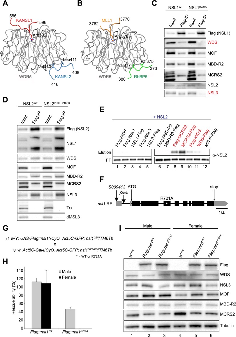
Figure 3.
The in vivo function of the NSL1–WDS–NSL2 interactions. (A,B) Comparison of the crystal structures of human ternary KANSL1–WDR5–KANSL2 (A) and MLL1–WDR5–RbBP5 (Protein Data Bank [PDB] code 3P4F) (B) complexes. The two complexes were superimposed using WDR5. (C) Flag immunoprecipitation of wild-type (WT) NSL1 and NSL1R721A in SL-2 cells. Both wild-type nsl1 and nsl1R721A were transiently transfected into SL-2 cells, and immunoprecipitation was performed using Flag-M2 resin. Antibodies used for Western blot analysis are indicated. (D) Flag immunoprecipitation of wild-type (WT) NSL2 and NSL2I160E–V162D in stably expressing SL-2 cells. Both wild-type nsl2 and nsl2I160E–V162D were transfected into SL-2 cells, and stably expressing cells were selected. Immunoprecipitation was performed using Flag-M2 resin. Antibodies used for Western blot analysis are indicated. (E) Western blot analysis of Flag pull-down assays using untagged NSL2 as the prey and either N-terminal or C-terminal 3xFlag-tagged NSLs as bait. (FT) Flowthrough sample. Interacting proteins are highlighted in red. The eluted prey proteins are shown in Supplemental Figure S1D. (F) A schematic representation of the structure of the RE transcript, encoded by nsl1/CG4699, used in this study. Exons (black bars), introns (black lines), and untranslated regions (UTRs) (gray bars) are shown. The ATG start and the STOP codon are indicated as well as the positions of the transposon insertions (black arrows). The site of the introduced mutation disrupting the NSL1 binding to WDS is marked by a white asterisk. The protein alteration is given at the top. (G) Details of the crosses used in the rescue experiments. (H) Relative percentage of adult male (gray bars) and adult female (black bars) viability upon ectopic expression of wild-type nsl1 and mutant nsl1R721A in the absence of endogenous NSL1 at 25°C. The non-CyO; Tb siblings in which endogenous nsl1 is expressed were used as internal controls and scored as 100% viable. The detail of the fly crosses is given in G. The error bars represent standard deviations of three independent crosses. (I) Western blot analysis of proteins extracted from third instar male and female larvae expressing either wild-type or mutant (R721A) NSL1 in the absence of endogenous NSL1. w1118 larvae were used as a wild-type control. Antibodies used for the Western blot analysis are indicated. Tubulin was used as a loading control.

