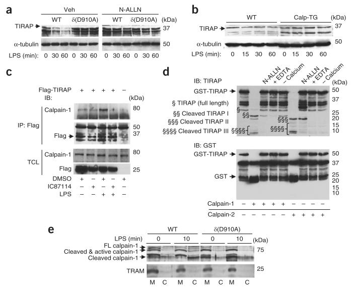
Figure 4.
‘Licensing’ of calpain-induced TIRAP proteolysis by p110δ in BMDCs and J774 macrophages. (a) Immunoblot analysis of TIRAP and α-tubulin in lysates of wild-type and δ(D910A) BMDCs pretreated for 1 h with vehicle or N-ALLN (50 μM) and then activated for 0–60 min (below lanes) with LPS (100 ng/ml). (b) Immunoblot analysis of TIRAP and α-tubulin in lysates of wild-type BMDCs and BMDCs with transgenic expression of calpastatin (calp-TG), treated for 0–60 min (below lanes) with LPS. (c) Immunoprecipitation and immunoblot analysis of J774 macrophages left untransfected (−) or transfected for 36 h to express Flag-tagged TIRAP (+), then pretreated for 1 h with vehicle (DMSO) or IC87114 (1 μM) and then left unstimulated or stimulated for 10 min with LPS (1 μg/ml), assessed with anti-Flag or anti-calpain-1 (above), along with immunoblot analysis of total cell lysates (TCL; below). (d) Immunoblot analysis of recombinant GST-TIRAP incubated with calpain-1 or calpain-2 (below lanes) in various assay conditions (above lanes). §, full-length TIRAP; §§, §§§ and §§§§, TIRAP cleavage products. (e) Immunoblot analysis of wild-type and δ(D910A) BMDCs left unstimulated (0 min) or stimulated for 10 min with LPS (100 ng/ml), followed by subcellular fractionation of membrane and cytosol. FL, full-length. Data are from one experiment representative of three with two to three mice per group (a,b) or one experiment representative of three (c–e).

