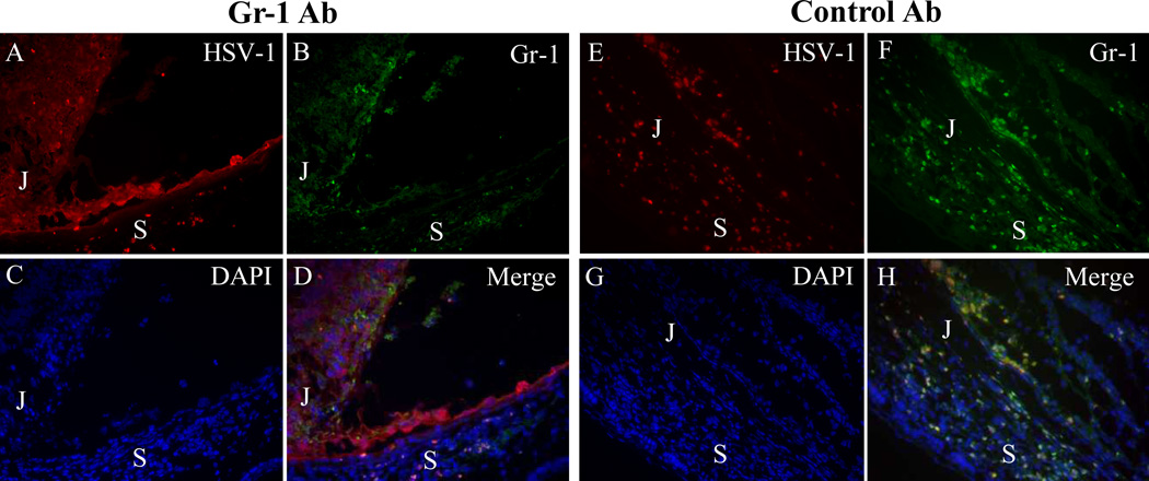
Figure 3.
Photomicrographs showing that neutrophil depletion correlated with increased HSV-1 infection of the parenchyma of the anterior segment of the inoculated eye at day 5 pi. (A–D) In Gr-1–treated mice, HSV-1–infected cells were observed at day 5 pi in the ciliary body, in RPE cells and in some peripheral retinal cells; a few Gr-1+ neutrophils were also seen. (E–H) Diffuse HSV-1 infection of the ciliary body and sclera with massive infiltration of Gr-1+ neutrophils was observed in the injected eyes of control antibody–treated mice. In control mice, most of the Gr-1+ cells were also HSV-1+ (compare D with H). (A, E) HSV-1 staining; (B, F) Gr-1 staining; (C, G) DAPI; (D, H) merged images. S, sclera; J, junction of ciliary body and pars plana.

