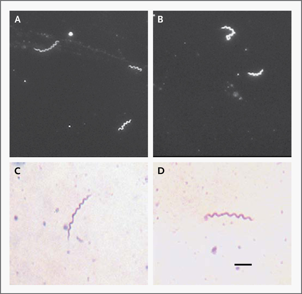
Figure 1. Morphologic Features of Spirochetes Detected in Cerebrospinal Fluid.
Panels A and B show the spirochetes as viewed with the use of dark-field microscopy. Panels C and D show the spirochetes as viewed with the use of bright-field microscopy, with Giemsa staining and a pH of 7.0. The bar indicates 2 µm.

