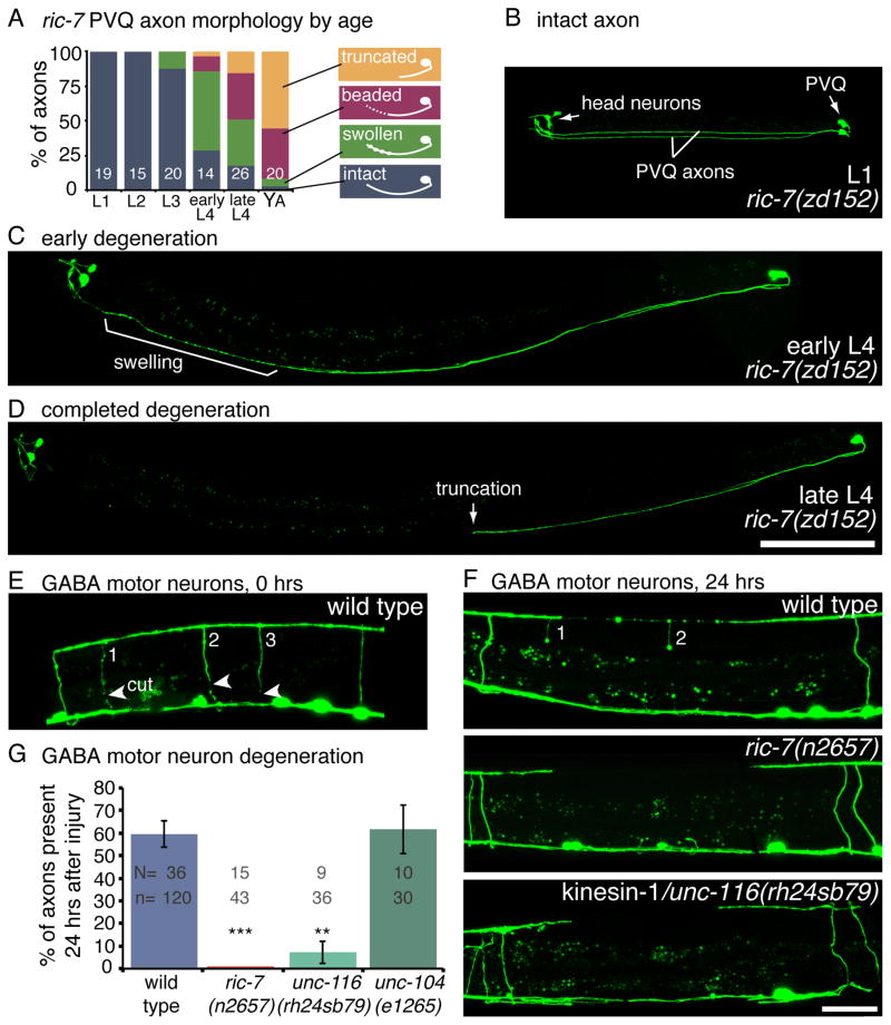
Figure 3. Axon degeneration is enhanced in the absence of mitochondria.
(A) PVQ axons spontaneously degenerate in ric-7 mutants. The degenerating axons proceed through the following morpholigies; swollen, beaded, and finally truncated. The percentage of axons with a given morphology is depicted for each life stage of ric-7(zd152). The number of worms assayed is indicated. (B) In the first larval stage, PVQ axons are intact in ric-7(zd152) mutants. GFP is expressed in PVQ and head neurons (ASH, ASI) under the sra-6 promoter. The two PVQ cell bodies are in the tail; each extends an axon along the ventral nerve cord to the head. (C) During the fourth larval stage, the distal portion of the axon degenerates, beginning with swellings. (D) PVQ axons are typically truncated, frequently near the vulva, by the end of the fourth larval stage. The proximal half of the axons and the cell bodies remain intact. Scale bar = 100 μm. See also Figure S3A–E. (E) GABA motor neuron axons degenerate following laser axotomy. Arrowheads point to the cut sites in the image taken immediately after axotomy. There are three severed axons (numbered). (F) After 24 hrs of recovery worms were reimaged and the presence or absence of severed axons was scored. The wild-type panel is the same worm from panel E. Two of the three axons exhibit swellings but are still present. Severed motor neuron axons completely degenerate in ric-7 and kinesin-1/unc-116 mutants. Scale bar = 25 μm. (G) The average percent of axons that are still present 24 hrs after axotomy +/− SEM. More axons degenerate in mutants lacking mitochondria compared to the wild type. The kinesin-3/unc-104 mutant, which has mitochondria throughout their axons, degenerates at wild-type levels. p < 0.0001 Kruskal-Wallis test with Dunn’s multiple comparisons to the wild type, *** p<0.001, **p<0.01. See also Figure S3F–K.

