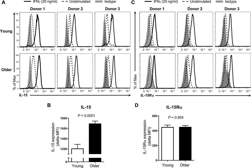
Figure 1. Older adults have increased surface expression of IL-15 on monocytes in response to IFN-γ compared to young adults.
Monocytes (CD14+CD16−) were negatively purified from PBMCs of young and older adults using a commercially available kit. Purified cells were stimulated for 16 hours with IFN-γ (20 ng/ml) or control (PBS). Cells were then stained with antibodies to IL-15 (A, B), IL-15Rα (C, D) or isotype control. Stained cells were analyzed on an LSRII® flow cytometer. (A) Representative histograms showing the induction of surface IL-15 expression on monocytes by IFN-γ. (B) Mean fluorescent intensity (MFI) of surface IL-15 expression on monocytes in response to IFN-γ in young and older adults. Delta MFI of IL-15 expression was obtained by subtracting MFI of IL-15 expression on IFN-γ-untreated monocytes from MFI of IL-15 expression on IFN-γ-treated cells. (C) Representative histograms showing the induction of IL-15Rα expression on monocytes by IFN-γ. (D) MFI of IL-15Rα expression on monocytes in response to IFN-γ in young and older adults. Delta MFI of IL-15Rα expression was obtained by subtracting MFI of IL-15Rα expression on IFN-γ-untreated monocytes from MFI of IL-15Rα expression on IFN-γ-treated cells. Data from 15 young and 19 old adults for B, and 16 young and 20 old adults for D. Bars and error bars indicate mean and standard error of the mean (SEM), respectively. P values were obtained by the Student’s t test.

