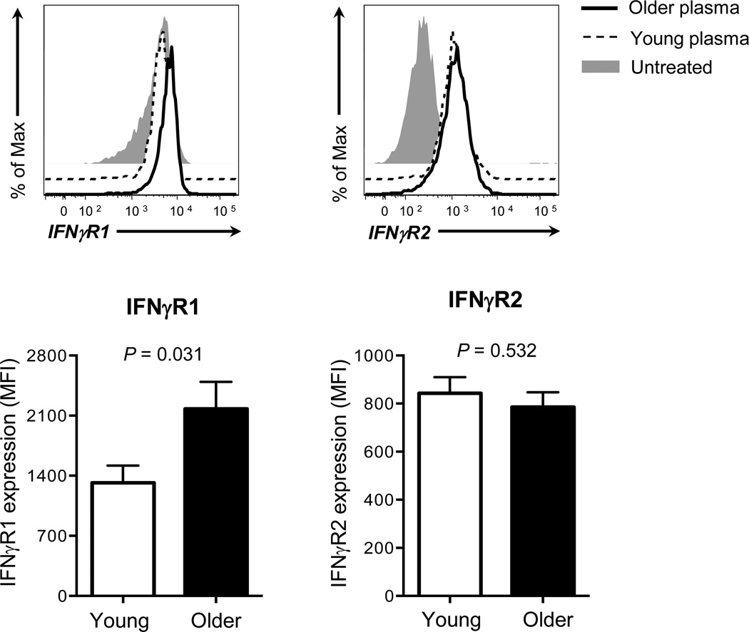
Figure 5. Human monocytes incubated with plasmas of young and older adults have increased expression of IFN-γR.
Monocytes (CD14+CD16−) were negatively purified from PBMCs of a young adult using a commercially available kit. Cells were incubated for 20 hours with or without plasmas from young and older adults (n = 12 and 12, respectively) (plasma to culture media volume, 1:1). Cells were then stained with Abs to IFN-γR1, R2 or isotype control followed by analysis on a flow cytometer. (A) Representative histograms showing the expression of IFN-γR1 on monocytes treated with or without plasmas. (B) Changes in mean fluorescent intensity (MFI) of IFN-γR1 expression on monocytes treated with plasmas of young and older adults. Delta MFI of IFN-γR1 expression was obtained by subtracting MFI of IFN-γR1 expression by plasma-untreated monocytes from MFI of IFN-γR1 expression by plasma-treated cells. (C) Representative histograms showing the expression of IFN-γR2 on monocytes treated with or without plasmas. (D) Changes in mean fluorescent intensity (MFI) of IFN-γR2 expression on monocytes treated with plasmas of young and older adults. Delta MFI of IFN-γR2 expression was obtained by subtracting MFI of IFN-γR2 expression by plasma-untreated monocytes from MFI of IFN-γR2 expression by plasma-treated cells. Bars and error bars indicate mean and standard error of the mean (SEM), respectively. Representative data from 2 independent experiments. P values were obtained by the Student’s t test.

