Figure 1.
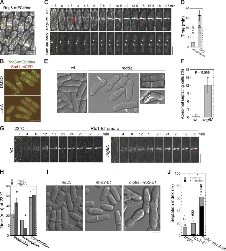
The novel protein Rng8 is involved in cytokinesis. (A) Rng8 localizes to the contractile ring (arrowheads), cables (arrows), and cytoplasmic puncta (asterisks). (B) Rng8 depends on actin filaments to localize. Cells were treated with DMSO or 100 µM Lat-A for 10 min at 25°C. (C) Time course (in minutes) of contractile ring formation in cells expressing Rng8-mEGFP Sad1-mCherry (Video 1). Time 0 marks SPB separation. The arrows and arrowhead indicate the Rng8 meshwork and the compact ring, respectively. In this and other figures, the cell boundary of some cells is marked with broken lines. (D) Time of appearance of Rng8 meshwork and the compact ring after SPB separation. Error bars indicate 1 SD. (E) Cytokinesis defects in rng8Δ cells. DIC images are shown. Arrows indicate examples of the aberrant septa. (F) Quantification of abnormal septating cells (see Materials and methods) as shown in E. n > 400 cells for each of three independent experiments. (G) Condensation of Rlc1 nodes into a compact ring (arrowheads) is delayed in rng8Δ at 23°C. Time 0 is the appearance of Rlc1 nodes. (H) Times of contractile ring assembly, maturation, and constriction in wt and rng8Δ cells (n > 25 cells for each) at 23°C. *, P < 0.001 compared with wt from two-tailed t test in this and other graphs. (I and J) Synthetic genetic interaction between rng8Δ and myo2-E1 and their septation indexes at 25°C. Bars, 5 µm.

