Figure 8.
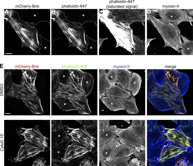
Bottleneck acts as a cross-linker/stabilizer of actin filaments. (A–D) Confocal images of HeLa cells expressing mCherry-Bnk (A) stained with phalloidin 647 (B and C) and myosin-II antibody (D) showing the colocalization between Bnk and actin. Nontransfected cells are marked with an asterisk. C shows the assembly of actin filaments in Bnk-overexpressing cells. In this panel, the phalloidin signal was adjusted to visualize actin in nontransfected cells. This resulted in the saturation of the phalloidin signal in the Bnk-transfected cell. Bar, 10 µm. (E) Confocal images of HeLa cells treated with cytochalasin D for 15 min, fixed, and stained with phalloidin 488 and myosin-II antibody. In Bnk-transfected cells, stress fibers do not depolymerize. DMSO-treated cells are shown as a control. Asterisks indicate nontransfected cells. Bar, 10 µm. See also Fig. S5.

