Figure 7.
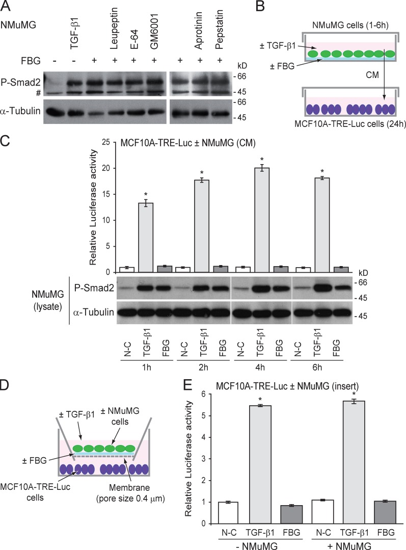
Mature TGF-β activated by the FBG domain of TNX is not diffusible, and its activation does not require any proteolytic event. (A) Phosphorylated Smad2 (P-Smad2) level in NMuMG cells seeded onto 222 pmol/cm2 FBG-coated (+) or uncoated (−) dishes and cultured for 3 h in the absence (vehicle) or presence of the indicated protease inhibitor (10 µM) or recombinant TGF-β1 (5 ng/ml). #, nonspecific band. (B) Culture of recipient MCF10A-TRE-Luc reporter cells with conditioned medium (CM) from donor NMuMG cells seeded onto a noncoated (N-C) or a 222 pmol/cm2 FBG-coated dish or treated with 5 ng/ml of mature TGF-β1 for 1–6 h. (C) Firefly luciferase activity of MCF10A-TRE-Luc cells cultured for 24 h with NMuMG cell–conditioned medium (top) and phospho-Smad2 levels in the donor NMuMG cells (bottom). White lines indicate that intervening lanes have been spliced out. (D) Co-culture assay of recipient MCF10A-TRE-Luc reporter cells (bottom well) with donor NMuMG cells (top well) seeded onto a porous membrane (pore size of 0.4 µm) coated or not coated with 222 pmol/cm2 of recombinant FBG domain or treated with 5 ng/ml of mature TGF-β1. (E) Firefly luciferase activity of MCF10A-TRE-Luc cells co-cultured for 24 h with or without NMuMG cells in the presence or absence of FBG domain or TGF-β1 as described in D. *, P < 0.05 versus uncoated condition. Error bars are means ± SD.

