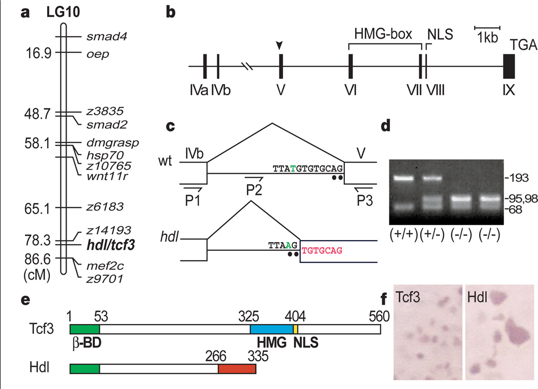
Figure 3.
Mapping of the hdl/tcf3 gene. a, Genetic map of linkage group 10 (LG10) showing position of hdl in relation to previously mapped genes and SSLP markers. b, Structure of the tcf3 gene. Arrowhead indicates the site of the hdl mutation. c, T-to-A transversion (green) produces an aberrant splice acceptor site (AG). The 7-bp insertion is indicated in red. d, Mse I digestion of the genomic PCR product (P2–P3), showing a polymorphism. e, Structure of Headless (Hdl). The truncated Hdl protein lacks the high mobility group (HMG) box DNA-binding motif as well as the nuclear localization signal (NLS). f, Subcellular localization of the wild-type Tcf3 and truncated Hdl proteins is changed from nuclear to cytoplasmic.

