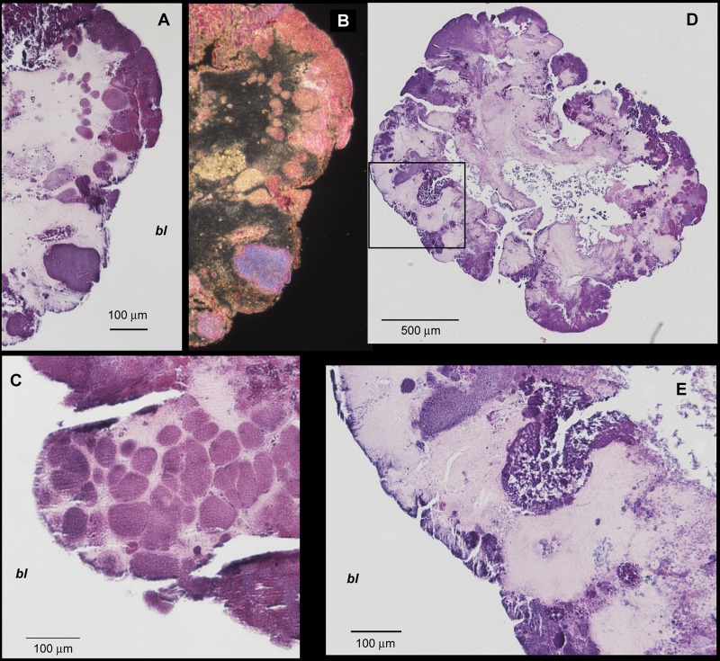
FIG 4.
Bright-field microimages from thin sections obtained from the center of granules and stained with H&E. The “bl” refers to bulk liquid. (A to C) Images show that various types of dense microcolonies (dark pink/purple) are embedded in a matrix of less dense cellular material (light pink). (D) Overview of an entire granule showing a highly irrigated inner structure. (E) Zoom image corresponding to the inset square in panel D showing colonies irrigated by the inner channels.

