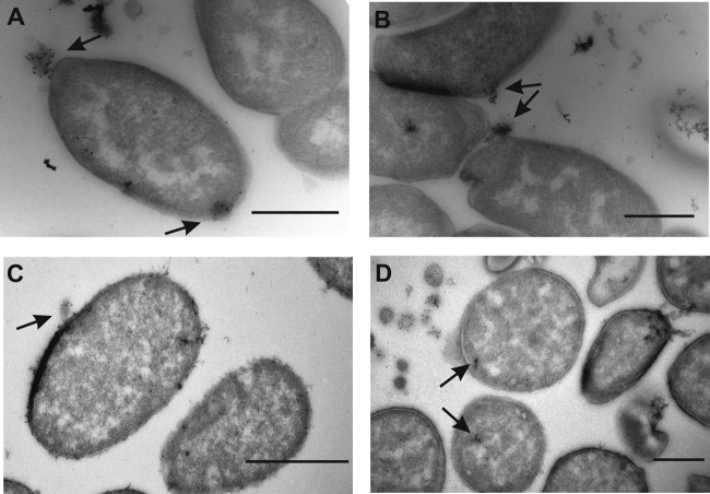
FIG 4.
TEM micrographs of FITC-labeled OMVs. (A, B) IEM micrographs of E. coli DH5α cells incubated with FITC-labeled OMVs derived from A. baylyi. The arrows point to the attached or internalized OMVs coupled to gold particles (5 nm) at exposure time t0 in panel A and at exposure time t1 in panel B. (C, D) Same as described for panels A and B but with A. baylyi JV26 cells. Bars, 500 nm.

