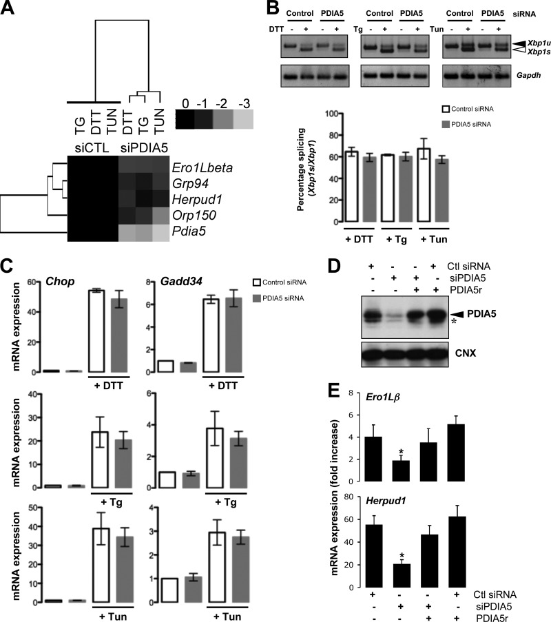
FIG 3.
Effects of PDIA5 silencing on ATF6α target genes and UPR signaling. (A) Heat map representation of the expression of ATF6α target genes upon silencing of PDIA5 using siRNA (25 nM) in HeLa cells. Forty-eight hours after transfection, cells were treated with 1 mM DTT, 500 nM Tg, or 5 μg/ml Tun for 2 h. Total RNA was isolated and analyzed by qPCR using specific primers for ATF6α target genes (Ero1Lβ, Grp94, Herpud1, and Orp150). Expression of each mRNA was normalized to that of Gapdh mRNA. (B) HeLa cells were transfected with siRNA and treated with DTT, Tg, or Tun, and the splicing of Xbp1 mRNA was evaluated using reverse transcription-PCR (RT-PCR). (C) RNA was extracted from control or PDIA5-silenced and ER-stressed HeLa cells and analyzed by qPCR using the specific primers Gadd34, Chop, and Gapdh. Data for qPCR are the mean ± standard deviation (SD) from three independent experiments. (D) HeLa cells were transfected with siRNA (control [Ctl] or PDIA5) and further transfected with pcDNA3-PDIA5r or not. Forty-eight hours later, lysates were analyzed by immunoblotting using either anti-PDIA5 or anti-CNX antibodies. The arrowhead shows PDIA5, and the asterisk indicates a nonspecific band. (E) Cells transfected as for panel D were then treated with 1 mM DTT for 2 h or not treated. RNA was then extracted, and the expression of Ero1Lβ and Herpud1 was evaluated by qPCR. Data are the average from three independent experiments ± SEM. *, P < 0.05.

