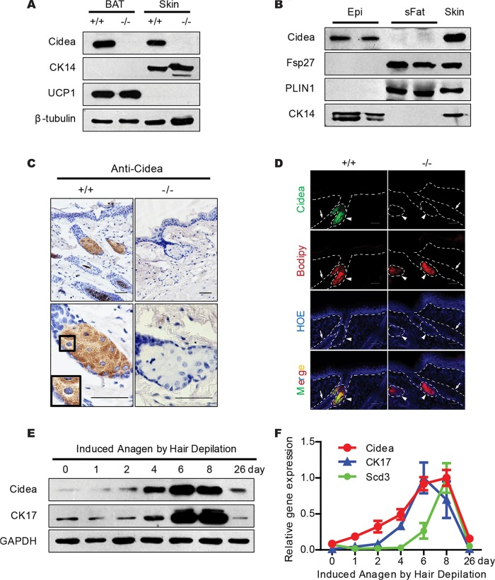
FIG 2.
Sebaceous-gland-specific and hair cycle-dependent expression of Cidea. (A) Western blot analysis showing the expression of Cidea protein in full-thickness mouse skin. β-Tubulin was used as a loading control; CK14 is a marker for skin; UCP1 is a marker for BAT. (B) Western blot analysis showing expression of Cidea in mouse skin epidermis (Epi) and of Fsp27 in subcutaneous fat (sFat). Epidermis was separated by incubating tail skin in 1.2 U/ml dispase. PLIN1 is a marker for subcutaneous white fat; CK14 is a marker for epidermis. (C) Immunohistochemical (IHC) staining of paraffin sections of skin from wild-type (+/+) and Cidea−/− (−/−) mice with Cidea antibody. Scale bar, 50 μm. (D) Immunofluorecence (IF) staining of frozen skin sections from wild-type and Cidea−/− mice using Cidea antibody. Neutral lipids were stained with Bodipy, and the nucleus was stained with Hoechst (HOE). Hair follicles are indicated by arrows and sebaceous glands by arrowheads. Scale bar, 50 μm. (E and F) Levels of Cidea protein (E) and mRNA (F) during induced anagen by hair depilation. GAPDH was used as a loading control; Scd3 and CK17 are markers for hair cycles. n = 5 for each indicated time point in panel F. Bars show means ± SEM.

