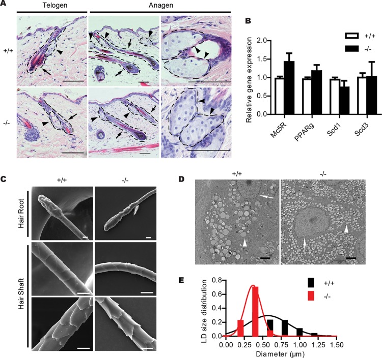
FIG 3.
Accumulation of smaller LDs in the sebaceous gland of Cidea−/− mice. (A) H&E staining of telogen and anagen skin sections from wild-type (+/+) and Cidea−/− (−/−) mice. Hair follicles are indicated by arrows and sebaceous glands by arrowheads. Scale bar, 100 μm. (B) Relative mRNA levels of sebaceous-gland-specific proteins (Mc5R, PPARγ, Scd1, and Scd3); n = 4 for each genotype. Bars represent means ± SEM. (C) Scanning electron microscopic images of hairs from wild-type mice (+/+) and Cidea−/− mice (−/−). No obvious differences can be seen in hair roots and hair shafts. Scale bar, 10 μm. (D) Electron microscopic (EM) images of skin sections from wild-type (+/+) and Cidea−/− (−/−) mice. Nuclei are indicated by arrows, and LDs are indicated by arrowheads. Scale bar, 2 μm. (E) Lipid droplet (LD) size distribution. n = 4 for each genotype. Diameters of over 570 LDs for each genotype were measured.

